Vision and Eye Diagram How We See
Ciliary body. The part of the eye that produces aqueous humor. Cornea. The clear, dome-shaped surface that covers the front of the eye. Iris. The colored part of the eye. The iris is partly responsible for regulating the amount of light permitted to enter the eye. Lens (also called crystalline lens).
:max_bytes(150000):strip_icc()/eye-conjunctiva-871453538-5a26c6ad22fa3a0037d5edad.jpg)
How the Human Eye Works (Structure and Function)
The iris is a flat, thin, ring-shaped structure sticking into the anterior chamber. This is the part that identifies a person's eye colour. The iris contains both circular muscles going around the pupil and radial muscles radiating toward the pupil. When the circular muscles contract, they make the pupil smaller.

Can We Grow New Eyes?
Aqueous humor - the clear, watery fluid inside the eye. It provides nutrients to the eye. Astigmatism - a condition in which the lens is warped, causing images not to focus properly on the retina. Binocular vision - the coordinated use of two eyes which gives the ability to see the world in three dimensions - 3D. Cones - cells the in the retina that sense color.

Human Eye Anatomy, Structure and Function
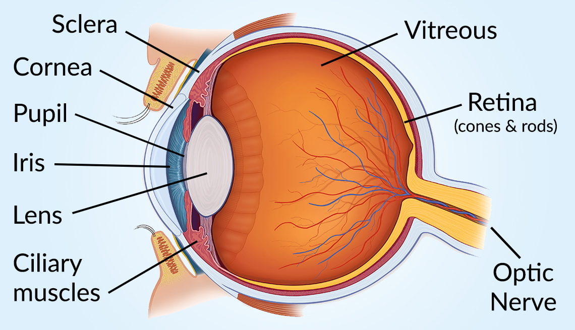
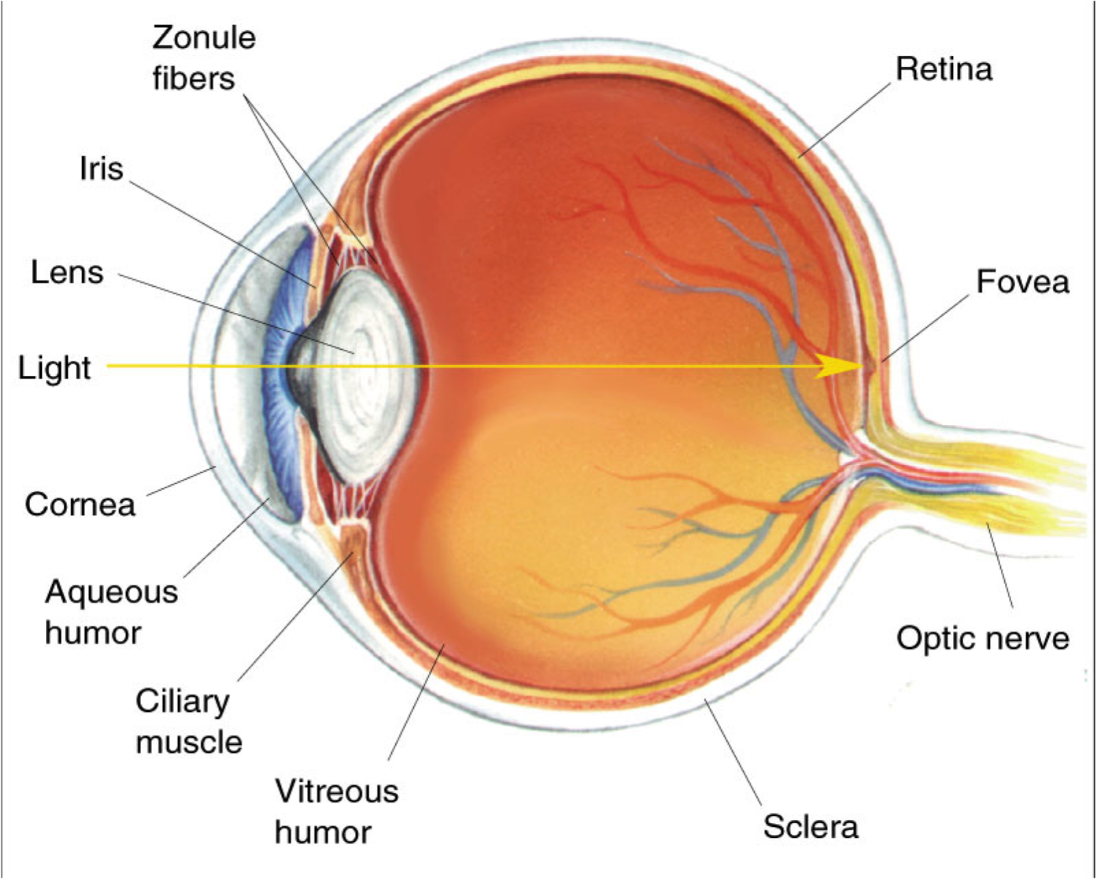
Behind the anterior chamber is the eye's iris (the colored part of the eye) and the dark hole in the middle called the pupil. Muscles in the iris dilate (widen) or constrict (narrow) the pupil to control the amount of light reaching the back of the eye. Directly behind the pupil sits the lens. The lens focuses light toward the back of the eye.
Human Eye Anatomy, parts and structure Online Biology Notes
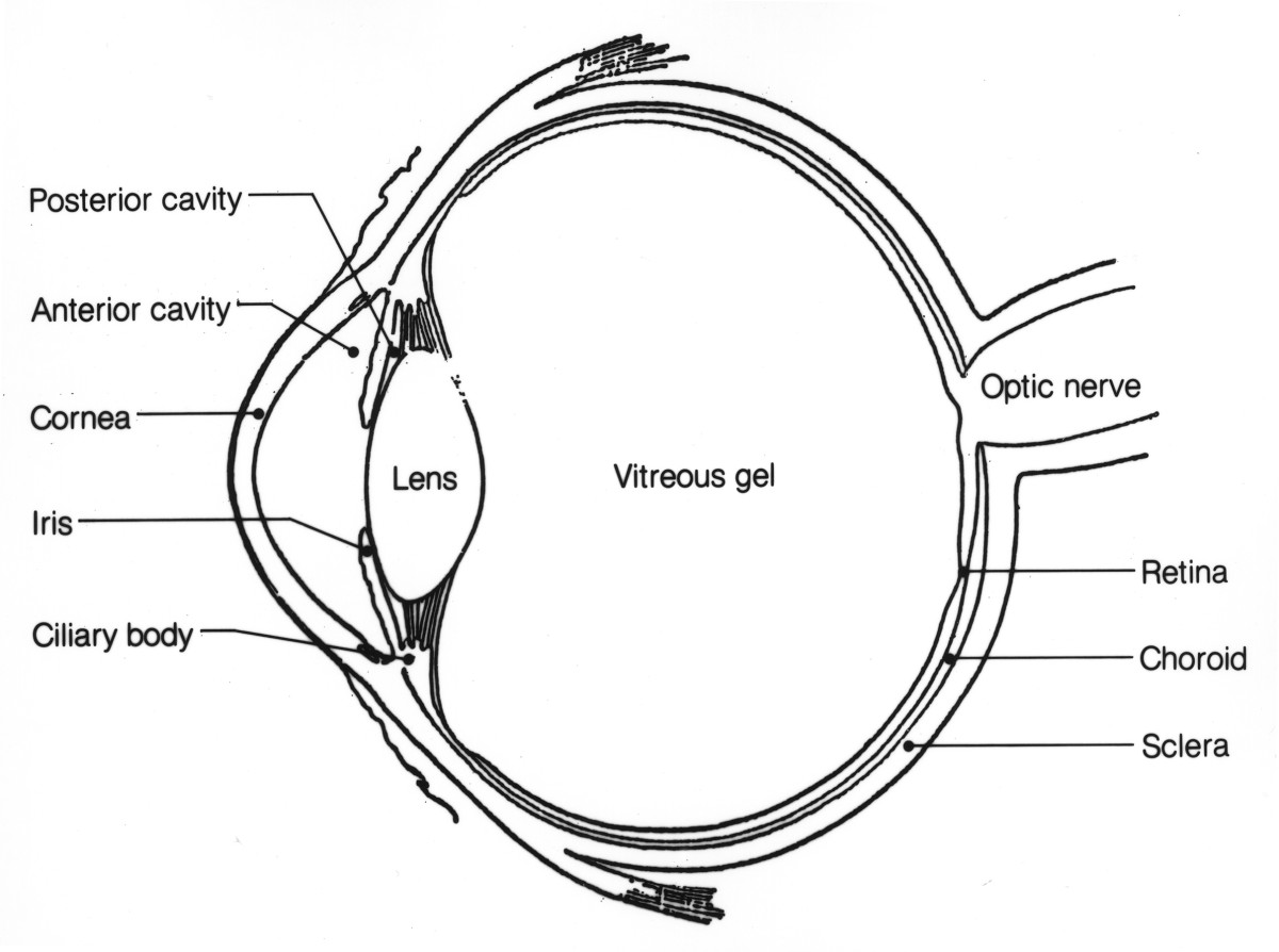
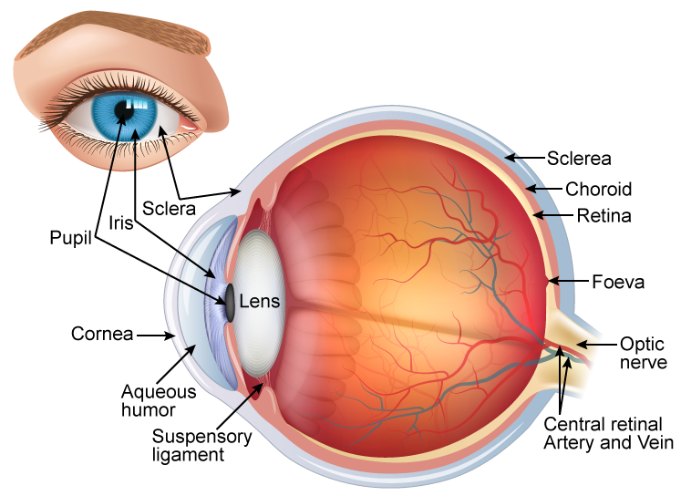
Take a look at the diagram of the eyeball above. Here you can see all of the main structures in this area. Spend some time reviewing the name and location of each one, then try to label the eye yourself - without peeking! - using the eye diagram (blank) below. Unlabeled diagram of the eye. Click below to download our free unlabeled diagram of.

OUR EYES WORK LIKE CAMERA’S! Discovery Eye Foundation
Pads of fat and the surrounding bones of the skull protect them. The eye has several major components: the cornea, pupil, lens, iris, retina, and sclera. These work together to capture an image.
Anatomy of the Eye Human Eye Anatomy Owlcation
Light is focused primarily by the cornea - the clear front surface of the eye, which acts like a camera lens. The iris (colored part) of the eye functions like the diaphragm of a camera, controlling the amount of light reaching the retina by automatically adjusting the size of the pupil (aperture). The eye's crystalline lens is located.
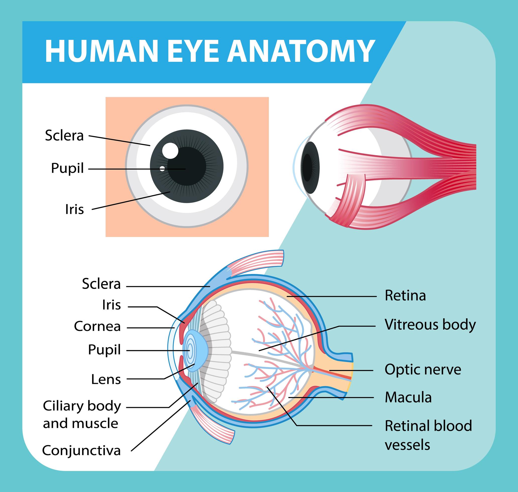
Diagram of human eye anatomy with label 1848847 Vector Art at Vecteezy
Anatomy of the Human Eye. Eyes are one of the most important organs of the body. A healthy pair of eyes means a clear vision, which plays a major role in day-to-day life and quality of experiences.
/GettyImages-695204442-b9320f82932c49bcac765167b95f4af6.jpg)
Structure and Function of the Human Eye
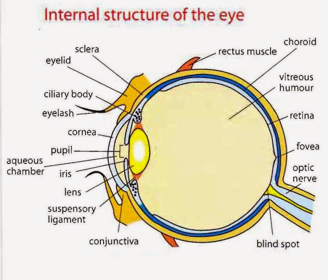
Structure of Human Eye. A human eye is roughly 2.3 cm in diameter and is almost a spherical ball filled with some fluid. It consists of the following parts: Sclera: It is the outer covering, a protective tough white layer called the sclera (white part of the eye). Cornea: The front transparent part of the sclera is called the cornea.

Eye Anatomy
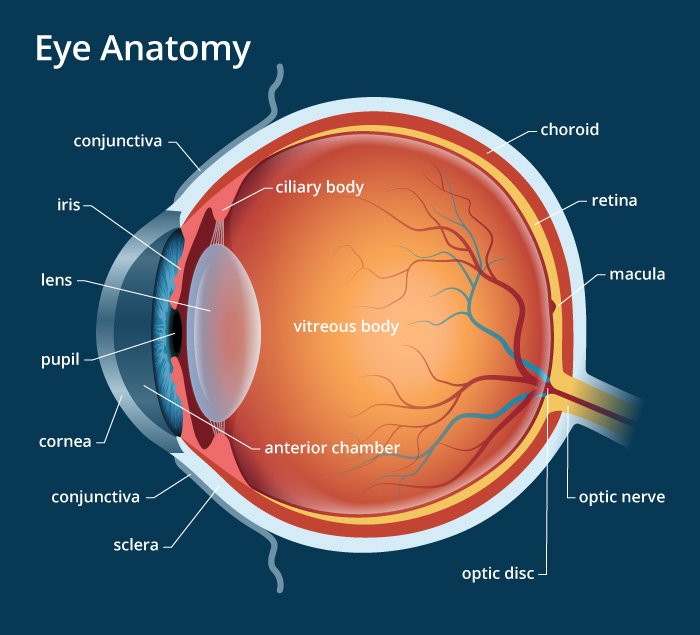
A brief description of the eye along with a well-labelled diagram is given below for reference. Well-Labelled Diagram of Eye. The anterior chamber of the eye is the space between the cornea and the iris and is filled with a lubricating fluid, aqueous humour. The vascular layer of the eye, known as the choroid contains the connective tissue.
HUMAN EYE (STRUCTURE, IMAGE FORMATION AND DIFFERENCE BETWEEN RODS AND CONES) « SimpleBiology
Labelling the eye. Use this interactive to label different parts of the human eye. Drag and drop the text labels onto the boxes next to the diagram. Selecting or hovering over a box will highlight each area in the diagram. The human eye has several structures that enable entering light energy to be converted to electrochemical energy.
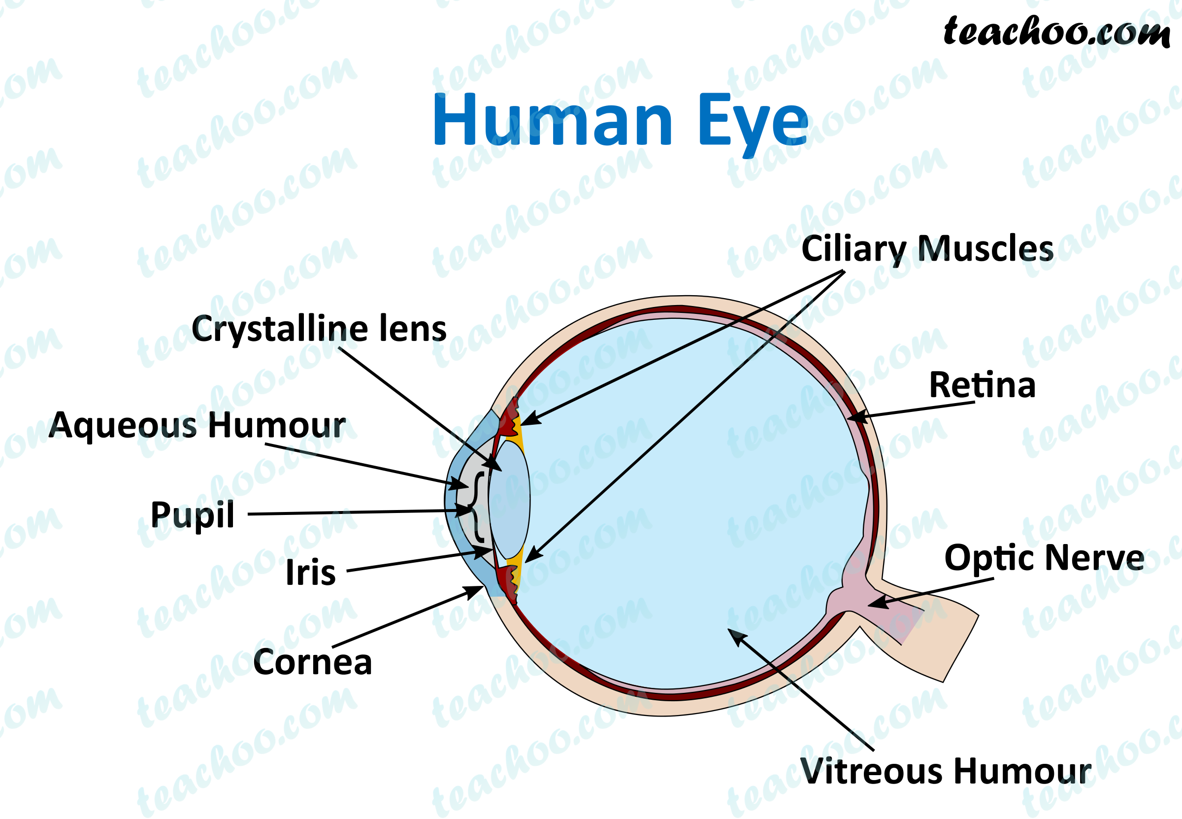
Human Eye Different Parts and their functions Class 10 Teachoo
The External Structure of an Eye. Sclera: It is a white visible portion. It is made up of dense connective tissue and protects the inner parts. Conjunctiva: It lines the sclera and is made up of stratified squamous epithelium. It keeps our eyes moist and clear and provides lubrication by secreting mucus and tears.

Human Eye Anatomy Parts of the Eye and Structure of the Human Eye
Cornea: The clear, dome-shaped tissue covering the front of the eye. Fovea: A tiny pit located in the macula of the retina that provides the clearest vision of all. Iris: The colored part of the eye that controls the amount of light that enters the eye by changing the size of the pupil. Lens: A crystalline structure located just behind the iris.
Eye Diagram Cliparts.co
Download. English: Parts of the Eye (PDF 603.5 KB) Spanish: Las partes del ojo (PDF 897.7 KB) Check out this fact sheet to see a labeled diagram of the eye and learn about the different parts of the eye.
draw a neat and labelled diagram of structure of the human eye slwbyx77 Science
Iris: The iris is the colored part of the eye that regulates the amount of light entering the eye. Lens: The lens is a clear part of the eye behind the iris that helps to focus light, or an image, on the retina. Macula: The macula is the small, sensitive area of the retina that gives central vision. It is located in the center of the retina.

3 Anatomy Surrounding the Eye OpticianWorks Online Optician Training Human anatomy and
The inferior rectus: Attaches to the bottom of the eye and allows downward eye movement. The medial rectus: Attaches to the side of the eye adjacent to the nose and helps the eyes to shift inwards towards the nose. The lateral rectus: Attaches to the outer side of the eyes and moves the eyes toward the temples.